Solving the mystery of deformed beaks
By Jack Dumbacher
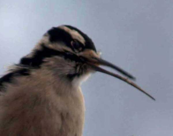
The second article suggested that the disease wasn’t limited to just a couple species, and also showed that it appeared to be spreading to both more birds and to more localities. The disorder was found in chickadees, nuthatches, crows, jays, woodpeckers, hawks – and these are just the easy-to-see birds that tend to hang out at people’s feeders or urban parks. The articles were partially a call to arms and a plea for more information from citizen birders and other scientists to recognize and help track the disease.
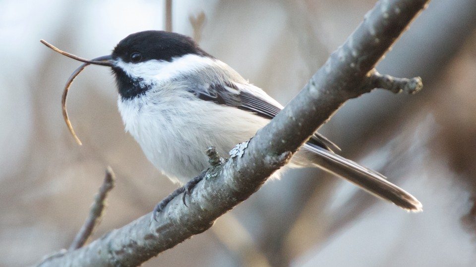
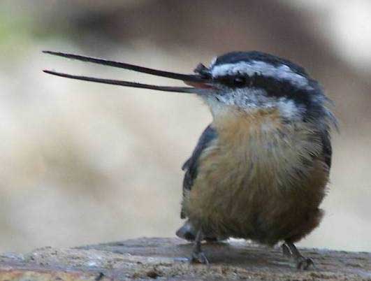
Despite investigating several possible causes, the authors were unable to determine what caused the beak deformities. They looked at known viruses, bacterial infections, fungus, mites – even potential environmental toxins. But no smoking gun.
At the same time, I was starting a collaboration with Joe Derisi, who is a virus researcher at University of California San Francisco and clever virus hunter (among other things). He had some fancy lab equipment and techniques for finding viruses. One tool was a virus microarray chip — their virochip — a small glass microscope slide with tens of thousands of short sequences that are made to match a portion of the genome of virtually every known virus. By putting DNA from a sick person (or bird) onto the microarray, a DNA match would light up and indicate a virus present, and potentially which one. We thought it would be fun to see if his virochip could identify a bird virus.
So I quickly contacted the key investigators, Colleen Handel and Caroline van Hemert from the USGS labs in Alaska. They were keen to try anything, and consented to send some samples taken from sick birds. We tried the virochip, and got some hits that we followed up, but answers weren’t quickly forthcoming. One challenge was that bird DNA was very different from human DNA, so we didn’t know what the background virochip pattern would look like when normal bird DNA was run on the chip. But it did suggest some leads that we followed up – one that led to the discovery of a new tiny circular virus (3). But after following up on this new virus, we decided that it probably wasn’t related to the disease.
But by then, we were hooked and invested in finding the cause. Derisi lab members were experimenting with another technique that we wanted to try. They were using new whole-genome sequencing techniques to look for viruses. It worked something like this:
Take a sample of blood or a mouth or cloacal swab from a sick bird, then extract DNA and RNA from the sample. Then shear up the DNA and RNA into billions of fragments about 400-500 bases long. Then use new Illumina genome analyzers to read the sequences of literally hundreds of millions of these fragments. Then use a computer to look for pieces that match (or nearly match) any known virus genomes. Finally, starting with a piece that matches a virus, try to build longer and longer sequences by searching the sequence dataset for additional sequences that overlap the starting sequence, and then continue extending as far as possible. Once you get these extended pieces of DNA sequence, some will be big enough to reliably determine what they match — such as the bird’s genome, or a viral genome, or bacterial genome, and so on.
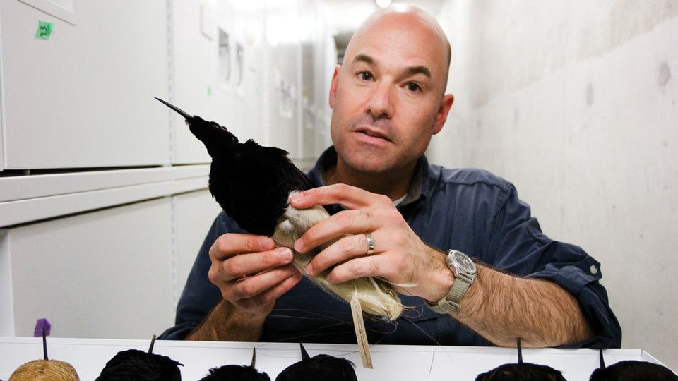
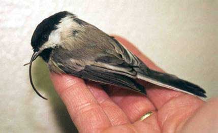
Around this point, we got a post-doctoral researcher, Maxine Zylberberg, to help with the virus work, and we set to work in the lab. We got additional Alaskan samples from Colleen and Caroline, and Maxine also added samples from some sick hawks from California. A team from Illumina generously offered to provide an entire flow cell of sequence data (amounting to nearly a billion short sequences, worth several thousand dollars at the time!!!) for the project. Before long, Maxine and Joe and I were looking through gene fragment assemblies that Maxine had produced, and two of the fragments provided a hit to a distantly-related picornavirus known from turkeys. Although far fewer than 50% of the amino acids were identical, the new sequence could be classified as a picornavirus by the genome structure, gene order and content. If you don’t speak virus, this all just means that this new virus was very distantly-related to any known virus, but it was a virus for sure. And Maxine used some other cool sequencing techniques to sequence the rest of the virus genome from the bird samples, finally nailing the ID of this new virus.
What we didn’t know at the time was that identifying this virus was the easy part. Maxine then worked for a couple years trying to definitively link this virus to the disease. She was able to screen lots of samples – and the birds with the disease usually tested positive for the virus (19 out of 19 Black-capped Chickadees), and healthy birds usually tested negative (7 out of 9 healthy chickadees were negative.) But this isn’t enough to prove that the virus CAUSED the disease. She tried to culture the virus in the lab, which turned out to be far from easy. And without cultures, it was impossible to do so-called challenge experiments where healthy birds are exposed to the culture to see if the disease can be spread. She additionally screened other bird species, and she was able to test various tissues to see where the virus hangs out. She named the new virus “Poecivirus” after the Black-capped Chickadee (Poecile atricapillus) that it came from. And Maxine did a lot of other cool stuff – all of which is reported in our recent paper (4).
During the time we were doing this work, cases of AKD were documented throughout Canada and the lower 48 states, and even a couple cases were documented in UK and Europe. The disease continues to spread in Alaska. Even if this new virus is the cause of the disease, we do not know whether it is spread directly from bird to bird, or through the environment by contaminated water or food (like bird feeders) or whether it is spread by a vector such as mosquitos. There is still much to learn about how serious the threat is to birds worldwide and how quickly it is spreading.
So now, with a viable candidate for the cause of AKD – what is next? There is a role for every birder out there – especially those who take photos. You can submit information about cases and photos of sick birds to USGS at their AKD website, or directly on their reporting site. Alternatively, you can email photos and information directly via this email link. This will be most helpful as Colleen and her team continue to track the spread and severity of the AKD outbreak.
Bibliography:
- Hemert, C.V. and C.M. Handel. 2010. Beak deformities in northwestern crows: evidence of a multispecies epizootic. The Auk, 127(4): p. 746-751.
- Handel, C.M., L.M. Pajot, S.M. Matsuoka, C.V. Hemert, J. Terenzi, et al. 2010. Epizootic of beak deformities among wild birds in Alaska: an emerging disease in North America? The Auk, 127(4): p. 882-898.
- Hanna, Z.R., C. Runckel, J. Fuchs, J.L. DeRisi, D.P. Mindell, et al. 2015. Isolation of a Complete Circular Virus Genome Sequence from an Alaskan Black-Capped Chickadee (Poecile atricapillus) Gastrointestinal Tract Sample. Genome announcements, 3(5): p. 1-2. doi: 10.1128/genomeA.01081-15
- Zylberberg, M., C. Van Hemert, J.P. Dumbacher, C.M. Handel, T. Tihan, et al. 2016. Novel Picornavirus Associated with Avian Keratin Disorder in Alaskan Birds. mBio, 7(4): p. e00874-16. doi: 10.1128/mBio.00874-16
Also – click here to download our California Academy Press release, with more information.
And check out this article about the work, on the Wildlife Society Blog.
